Microscopy

Modern microscopy techniques are able to penetrate into tissue to provide depth sectioning and/or 3D information. Confocal Laser Scanning Microscopy (CLSM), Optical Coherence Tomography (OCT), and Two-Photon Microscopy (TPM) have been very useful for imaging the retina. There is interest in applying these techniques to brain imaging driven by the field of Optogenetics.

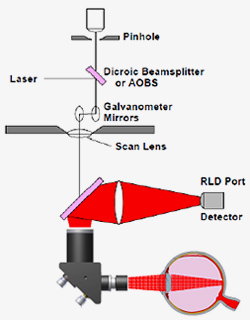
 

Modern microscopy techniques are able to penetrate into tissue to provide depth sectioning and/or 3D information. Confocal Laser Scanning Microscopy (CLSM), Optical Coherence Tomography (OCT), and Two-Photon Microscopy (TPM) have been very useful for imaging the retina. There is interest in applying these techniques to brain imaging driven by the field of Optogenetics. The ability to achieve high resolution imaging of deep tissue is limited by aberrations from the intervening layers of tissue.

Adaptive optics (AO) can enhance these techniques to improve imagery. Indeed AO has been incorporated by numerous researchers in the past several years to CLSM, OCT, and TPM with promising results.

Microscopy researchers are in the process of transferring astronomical AO techniques to the biological domain, including multi-dither coherent phasing, fluorescence guidestars, and multi-conjugate AO. Emphasis on speed is the next frontier as in-vivo adaptive-optics correction is desired.

AO microscopy tends to be performed with custom microscope systems on a breadboard laboratory environment. AOA Xinetics has designed, assembled and delivered an AO add-on unit to a commercially available two-photon microscope.

Schematic of light detection pathway in microscope

Prototype AO Optical Bench

Periscope Relay